
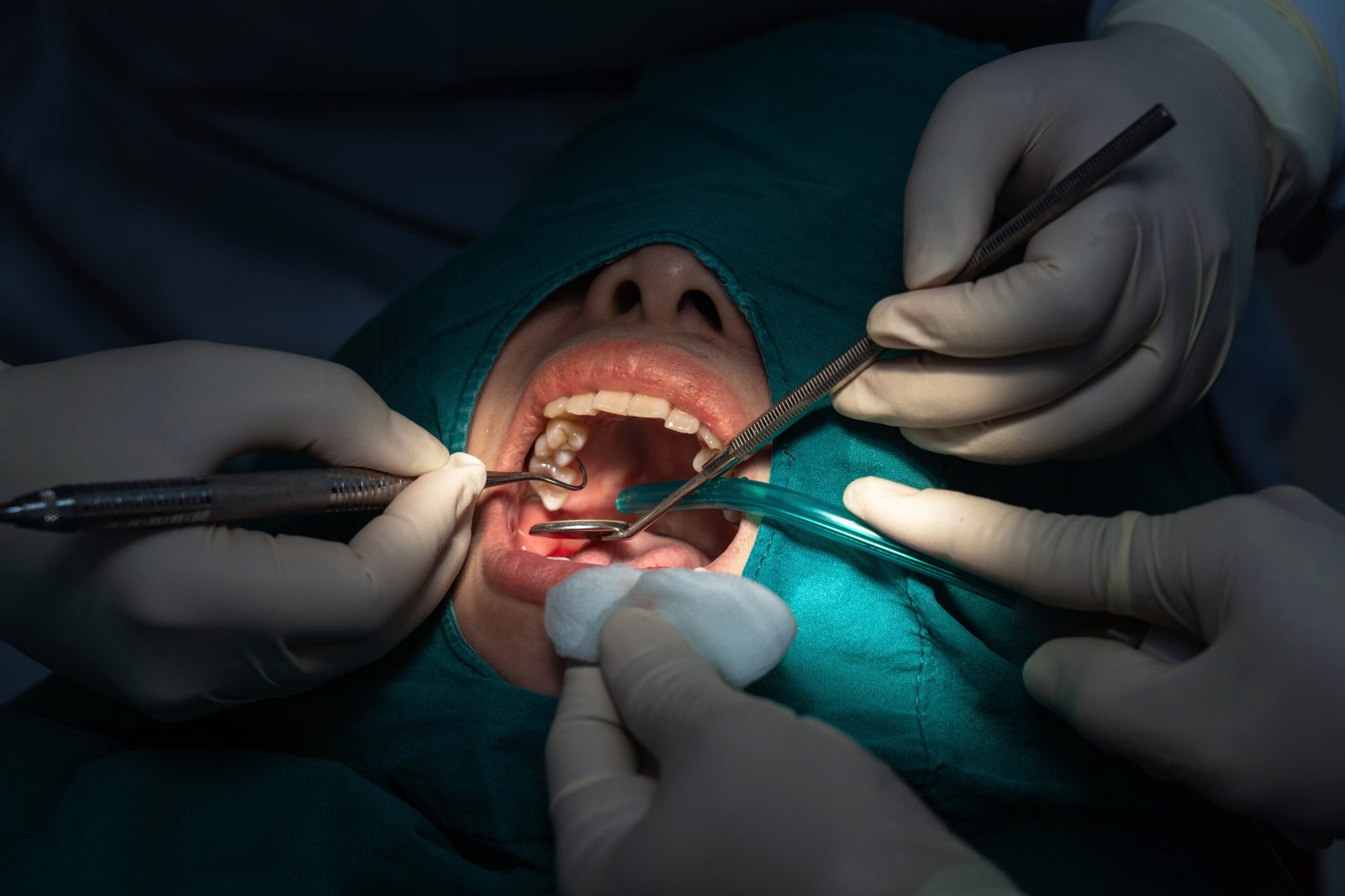
A wisdom tooth can look perfectly ordinary from the part you can see and still cause problems beneath the gum.
Another can look awkward on an X-ray yet remain quiet, stable, and appropriate to monitor.
That is why wisdom tooth removal should not be decided by one question alone:
“Is the tooth impacted?”
Impaction matters, but it is only the beginning.
Before recommending removal, the dentist needs to understand where the tooth sits, what it is doing to the tooth beside it, how its roots relate to nearby structures and whether there is evidence of disease around it.
The real question is not simply whether the wisdom tooth has enough space.
It is:
“What is this tooth doing now, and what does its position mean for treatment?”
A useful wisdom-tooth assessment should establish that.
“Impacted” Describes Position, Not the Whole Diagnosis
A wisdom tooth is described as impacted when it hasn’t erupted into a functional position.
It may be completely covered by gum or bone, partly erupted, tilted toward the neighboring tooth or positioned at another angle.
But the word impacted does not automatically tell you whether removal is necessary.
Some impacted teeth develop problems. Others may remain symptom-free and be monitored depending on their condition, position, and surrounding tissues.
That is why a clinical examination and appropriate imaging matter.
The dentist is not simply looking for an impacted tooth.
They are looking for the consequences of that position.
The Four Questions a Wisdom Tooth X-Ray Should Help Answer
A useful way to understand the assessment is through four questions:
Position → Neighbor → Roots → Disease
These four areas often tell the dentist far more than the simple label “impacted”.
1. Where Is the Wisdom Tooth Actually Positioned?
The first question is anatomical.
Is the tooth:
- fully erupted?
- partly erupted?
- completely impacted?
- angled toward the second molar?
- positioned horizontally?
- trapped beneath bone or gum?
Position influences both the likelihood of problems and the way treatment may need to be approached.
A fully erupted wisdom tooth that is healthy, functional, and easy to clean differs greatly from a partially erupted tooth that repeatedly traps food and becomes inflamed.
Likewise, a tooth lying horizontally against the molar in front presents a different situation from one that is upright but simply has not erupted.
That’s why you can’t assess an impacted wisdom tooth from symptoms alone.
The position provides context for everything that comes next.
2. What Is Happening to the Tooth Next to It?
This is one of the most important questions and one patients may not expect.
A wisdom tooth does not exist in isolation.
The second molar directly in front of it can sometimes be at greater risk.
Depending on position and access for cleaning, the area between the two teeth may become difficult to maintain. Dentists may look for evidence of:
- decay on the wisdom tooth
- decay on the neighboring molar
- gum problems behind the second molar
- pressure-related damage in selected cases
- food trapping and recurrent inflammation
That changes the decision considerably.
A wisdom tooth that causes little direct discomfort may still need attention if it compromises the adjacent healthy tooth.
So one of the most useful questions to ask during an assessment is:
“Is this wisdom tooth affecting the tooth in front of it?”
Protecting the second molar can sometimes be more important than focusing on the wisdom tooth itself.
3. What Do the Roots Look Like and What Is Nearby?
The roots matter because they help determine how straightforward or complex removal may be.
Dentists assess their number, shape, direction, and stage of development, along with their relationship to nearby anatomical structures.
For lower wisdom teeth, dentists may pay particular attention to the relationship between the roots and the nerve canal running through the lower jaw.
This does not mean that every lower wisdom tooth close to the nerve is unsafe to remove.
It means the relationship needs to be understood before planning treatment.
A standard dental X-ray or panoramic image may provide the necessary information often. Where the relationship is unclear, or greater three-dimensional detail could change management, additional imaging may be considered.
That is a clinical decision, not something every patient automatically needs.
This is also why surgical wisdom tooth removal cannot be planned responsibly from a photo of the tooth or a description of pain over the phone.
The anatomy matters.
4. Is There Evidence of Disease Around the Tooth?
Position alone is only part of the story.
The dentist also looks for evidence that the wisdom tooth or surrounding tissues are already unhealthy.
This may include:
- decay
- repeated gum inflammation around a partly erupted tooth
- infection
- damage to the adjacent tooth
- changes in the surrounding bone or tissues
- other clinically significant findings
This distinction matters because a tooth can be painless and still require investigation, while pain around the back of the jaw does not automatically prove that extraction is the correct treatment.
Symptoms tell the dentist where to look.
Examination and imaging help establish what is actually happening.
Pain Is a Clue, Not the Entire Decision
Wisdom tooth pain can be caused by several things.
A partially erupted wisdom tooth may develop inflammation in the gum around it. Food can become trapped. Decay may be present. A neighboring tooth may be the true source of pain.
The discomfort may also come and go.
That is why “it stopped hurting” does not necessarily mean the underlying problem has disappeared.
Simultaneously, not every episode of discomfort means the tooth must be removed immediately.
A proper assessment should identify the cause before moving to treatment.
The principle is simple:
Do not remove a tooth because of a label. Remove it when the clinical findings justify removal.
When Can a Wisdom Tooth Be Monitored Instead?
This is an important part of the conversation because not every wisdom tooth requires extraction.
A tooth may sometimes be monitored when it is healthy, symptom-free, not damaging nearby structures, and can be reviewed appropriately over time.
Monitoring does not mean forgetting about it.
The dentist may still recommend periodic examination and, where clinically appropriate, imaging to watch for changes.
The decision can also change later.
A tooth that is stable today may develop decay, gum problems or another complication in the future.
So the real decision is not simply:
Remove or ignore.
It may be:
Remove → Treat another problem → Monitor with review
That is a more accurate way to think about wisdom-tooth management.
When Does Removal Become More Likely?
The case for wisdom tooth extraction in Dubai patients becomes stronger when the tooth is linked to a clear clinical problem.
Examples include recurrent inflammation or infection, decay that cannot be managed predictably, damage to the adjacent molar, or another significant finding associated with the tooth.
Removal may also be considered as part of a wider treatment plan when there is a specific clinical reason.
The important part is that the recommendation should have an explanation behind it.
You should be able to ask:
“What problem are we solving by removing this tooth?”
Also, receive a clear answer.
Simple Extraction or Surgical Removal?
Not every wisdom tooth is removed in the same way.
A fully erupted tooth with accessible anatomy may be removed with a relatively straightforward extraction.
A tooth that is partly or fully trapped beneath gum or bone may require surgical access.
That can involve opening the gum and, where necessary, removing a controlled amount of surrounding bone or dividing the tooth into sections so it can be removed safely.
The exact technique depends on anatomy.
So the difference between a simple extraction and surgical wisdom tooth removal isn’t just how painful the tooth feels.
It largely depends on how the tooth is positioned and how accessible it is.
What Should You Ask Before Wisdom Tooth Removal?
You do not need to understand every detail on the X-ray.
But you should understand the treatment decision.
Useful questions include:
- What problem is this wisdom tooth causing right now?
- Is the tooth affecting the molar next to it?
- Is it fully erupted, partly erupted, or impacted?
- Is its root position relevant to the complexity of removal?
- Would this be a simple extraction or surgical removal?
- Is monitoring a reasonable alternative in my case?
These questions turn consent into an informed discussion rather than simply agreeing because an X-ray shows a tilted tooth.
What Happens After Removal?
Recovery depends on the tooth, the procedure, and the individual patient.
Your dentist should provide specific aftercare instructions, including guidance on oral hygiene, food, activity, and any prescribed medication.
Some temporary discomfort, swelling, or limited mouth opening can occur after wisdom-tooth surgery, particularly following more involved procedures.
Follow the instructions you are given and contact the dental clinic if something feels significantly worse than expected or if you are concerned about healing.
Consider aftercare part of the treatment plan, not an afterthought once the tooth is out.
When Should You Seek Prompt Evaluation for Wisdom Tooth Symptoms?
Arrange dental assessment if you develop persistent or recurring pain at the back of the mouth, swelling, difficulty cleaning around a partly erupted tooth, or repeated inflammation.
More urgent evaluation may be needed if there is:
- significant or spreading facial swelling
- fever or feeling systemically unwell
- severe worsening pain
- difficulty opening the mouth
- difficulty swallowing
- difficulty breathing
Difficulty breathing or significant swallowing problems require urgent medical attention.
When infection is suspected, waiting for the tooth to “settle down again” is not a reliable treatment strategy.
The Best Wisdom-Tooth Decision Starts With Anatomy, Not Assumptions
A wisdom tooth can be impacted without currently causing a problem.
It can be painless while affecting the adjacent tooth.
It can hurt even when extraction is not the first conclusion.
And two teeth producing almost identical symptoms can present disparate surgical considerations on imaging.
That is why good treatment planning asks more than:
“Is it impacted?”
It asks:
Where is it?
What is happening to the neighboring tooth?
What do the roots look like?
Is there disease around it?
Those answers determine whether the next step is treatment, removal, further investigation, or monitoring.
For patients in Business Bay and surrounding areas who need a wisdom-tooth assessment, White Swan Dental Clinic can examine the area, review appropriate imaging, and explain the available options according to the individual clinical findings.
FAQs
Does every impacted wisdom tooth need to be removed?
No. An impacted tooth may sometimes be monitored if it is healthy and not causing damage or disease. The decision depends on its position, surrounding tissues, symptoms, and clinical findings.
Can a wisdom tooth cause damage without pain?
Yes. Some problems can develop with limited symptoms, including decay or changes affecting the neighboring molar. This is why examination and appropriate imaging can be important.
Do I always need an X-ray before wisdom tooth removal?
Imaging is commonly used to assess the tooth, roots, and surrounding structures before extraction. The type of imaging required depends on the individual case and what information the dentist needs for treatment planning.
What is the difference between simple and surgical wisdom tooth extraction?
A simple extraction is generally possible when the tooth is erupted and accessible. Surgical removal may be required when the tooth is partly or completely trapped beneath gum or bone.
Can wisdom tooth pain go away without extraction?
Symptoms can sometimes settle temporarily, but that does not confirm that the underlying cause has resolved. A dental assessment is needed to determine whether treatment, monitoring, or removal is appropriate.